Helicobacter pylori detection and elimination
By: Dr. Natalia Loaiza Diaz
Medical Microbiologist, Clinical Pathology Leader. Laboratorio Clínico Hematológico S.A.S.
Published: 25 April 2023
How to diagnose H. pylori infection?
The first thing to do is to consult when symptoms of gastritis are present (pain or burning in the upper abdomen, accompanied or not by nausea, vomiting, a feeling of fullness or lack of appetite), when a family member or partner has been informed that he or she has the infection, or in cases of unexplained iron deficiency anemia or idiopathic thrombocytopenic purpura. 1,2. According to the physician's findings during the interview and physical examination, he/she will indicate the test he/she considers pertinent.
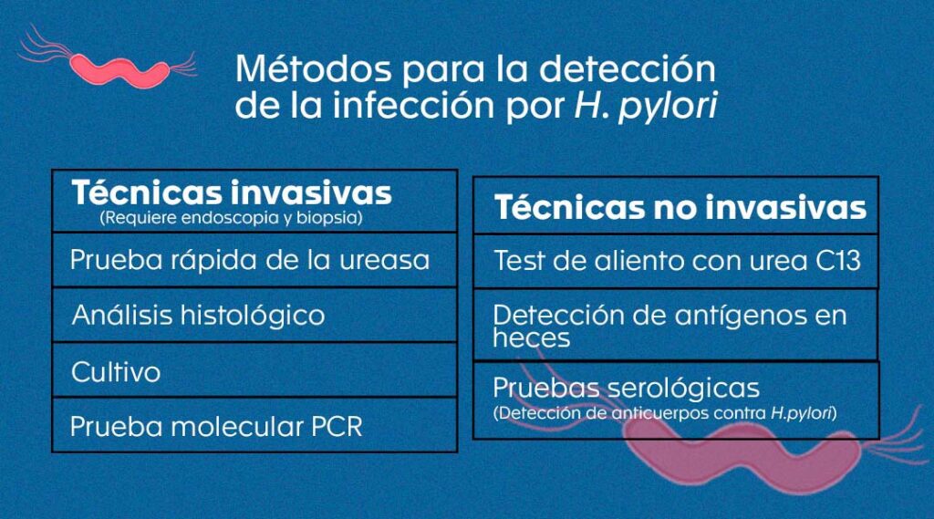
Currently, several types of tests are available in the field to approach the diagnosis of this infection. Some of them require gastric tissue obtained by biopsy during digestive endoscopy procedures; therefore, they are considered invasive. Others look for "clues" to the microorganism indirectly, i.e. without reaching the tissue, and are therefore considered non-invasive.
For the results of both groups of tests to be optimal and to avoid false results, it is essential that the patient stops taking drugs such as antibiotics (four weeks before) and proton pump inhibitors (two weeks before), except for the serum (blood) antibody detection tests, which do not require it. 3-9
Non-invasive tests:
- Urea breath test 3-6,8,10.
This test is considered the best among the non-invasive methods to make the diagnosis of H. pylori infection and is available at Hematologic. It is useful both for initial diagnosis and to confirm whether treatment was successful once it has been completed.
The test measures the activity of the urease enzyme produced by the bacteria to settle in the stomach. It consists of administering orally to the patient (on an empty stomach) a dilution containing a type of urea labeled with carbon 13 (13C-urea), a non-radioactive isotope of carbon. If H. pylori is present, its enzyme urease will hydrolyze or decompose the 13C-urea into ammonia and bicarbonate. The latter will be labeled with 13C and will pass into the circulation, travel to the lungs and exit on exhalation in the form of carbon dioxide labeled with 13C (13CO2).
Subsequently, a sample of exhaled air (or breath) is taken from the patient, and the 13CO2 released compared to that of a breath sample taken prior to ingesting the solution of 13C-urea solution, using specialized equipment that allows for the separation of 13 13CO2 from the CO2 normally exhaled.
- Detection of H. pylori antigen H. pylori in fecal material 3-5,8,9,11.
This is another non-invasive test, available at Hematológico through the network of allied laboratories. Like the previous one, it is useful for initial diagnosis and to check therapeutic success. For this test, techniques such as enzyme immunoassays or immunochromatography are used to indirectly determine the presence or absence of the microorganism in stool samples by means of antibodies directed against H. pylori contained in the test, which will specifically bind to the antigens of the bacterium and thus reveal whether it is present in the patient's sample.
- Detection of IgA, IgM and IgG antibodies against H. pylori 3-5,8,11.
This non-invasive technique is available at the Hematológico through allied laboratories and allows to know if the person has been in contact with the microorganism previously, since it measures immunoglobulin (Ig) antibodies of different classes (IgA, IgM and IgG) produced by the patient's immune response against H. pylori. The sample used is serum (blood) obtained by venipuncture.
The antibodies measured in this test cannot differentiate between a past and an active infection, nor does their value relate to the severity of the infection. Furthermore, since they can remain in the blood for more than one year, they should not be used to determine whether or not the treatment was successful.
Invasive tests:
To perform the procedure, the patient must first undergo an endoscopy procedure, which is usually performed under sedation. In this procedure, in addition to assessing the condition of the stomach mucosa with the aid of a camera, biopsies can be taken. What is recommended by the Sydney system, a standardized method used worldwide for the classification of gastritis and the reporting of endoscopic and biopsy findings, in its 1994 updated version, is to take five tissue fragments or biopsies, as follows: two from the antrum, two from the body, and one from the incisure. 12,13.
The aim is to increase the probability of detecting the microorganism and to detect premalignant lesions in a timely manner. 12,1312,13 because the greater the amount of tissue seen under the microscope or sown in culture, the greater the probability of observing H. pylori or obtaining its colonies.
- Histological examination1,3-6,11.
This study, available in the anatomic pathology laboratory of the Hematológico, is the most important of the analyses to be performed on samples obtained by invasive methods. It consists of submitting the gastric tissue, which must be in bottles with buffered formalin provided by the laboratory, to a series of processes to preserve its structure and stain it with hematoxylin-eosin, Giemsa and Warthin-Starry silver stains, and sometimes with immunohistochemical stains.
Once they are ready to be mounted, the stained slides are visualized under the light microscope to determine not only whether or not H. pylori is present in the tissue, but also active inflammation, changes in tissue architecture, cell shape and arrangement, among others, and finally to conclude whether or not there is infection, classify the gastritis and indicate whether there are changes associated with premalignant or malignant lesions.
- Urease rapid test 4,8.
It consists of subjecting a piece of a gastric biopsy to contact with a reagent containing urea and, using the presence of the urease enzyme of the microorganism in the sample, to appreciate a color change in the urea that is produced by the change in pH that is generated when the urease of H. pylori hydrolyzes urea into bicarbonate and ammonia. This test, available in the anatomic pathology laboratory of the Hematológico, has the advantage that the results are obtained quickly and can be used in the initial diagnosis, but not for follow-up treatment since a new biopsy would be necessary.
- Culture of Helicobacter pylori 1,4,11,14.
This method is considered the gold standard for making the diagnosis of infection. The test is offered in few centers since the characteristics of the microorganism require the use of special transport and culture media, and unusual procedures for processing, seeding of the samples and incubation of the culture media, to finally reach the point of value of this test which is the isolation of the bacteria, with the subsequent performance of antimicrobial susceptibility testing.
At Hematológico we directly culture gastric tissue biopsies taken by the endoscopist and placed in the transport medium previously provided by the laboratory. Ideally, their processing and seeding in the culture media is done the same day the samples are taken. Subsequently, they are incubated in an atmosphere containing higher humidity and CO2 than usual, and observed periodically, waiting to observe from the fifth day onwards, a small dew that indicates that H. pylori is growing.
Finally, starting from the colonies of the microorganism that have grown on the agar, tests are performed to confirm its identification and susceptibility to antimicrobials (antibiogram), which usually takes another three days. In this case, the bacterium is challenged against five antibiotics: amoxicillin, clarithromycin, metronidazole, levofloxacin and tetracycline, in order to determine in vitro whether it is sensitive or resistant to these agents.
- Molecular tests for the detection of H. pylori
Molecular biology techniques such as polymerase chain reaction (PCR) offer great advantages, as they are more sensitive than culture in detecting and identifying the microorganism in the sample, as well as some resistance genes, mainly for macrolides (such as clarithromycin) and fluoroquinolones (such as levofloxacin), and the result is obtained in a single day. 4,7,14,15.
Such tests can be applied directly on tissue fragments (gastric biopsies), on bacterial colonies grown in culture, even on stool samples. 1,4,8,111,4,8,11 However, in the country their availability is still scarce in clinical laboratories.
PCR and other techniques such as NGS (next-generation sequencing) are also used to study the presence of genes and mutations associated with increased virulence of the microorganism and greater severity of infection in certain cases and populations. 8,16
How can H. pylori infection be eliminated?
The treatment of this infection requires the combination of several drugs, such as proton pump inhibitors and antibiotics from different families (amoxicillin, clarithromycin or metronidazole), which must be taken for 14 days and continuously. This combination is called standard triple therapy and is recommended by international guidelines and the Colombian clinical practice guidelines for the diagnosis and treatment of H. pylori infection in adults, as the first line of treatment. 6,12,14
Pharmacological treatment is often selected empirically; however, keeping in mind that from the time the bacteria colonize the stomach until they trigger changes in gastric tissue and cancer, many years elapse 17and it is known that the microorganism has developed resistance to several of the antibiotics commonly used for its treatment, it is considered a good practice to perform culture or molecular tests to identify the bacteria and to know its susceptibility profile before defining the treatment. This, in order to choose the antibiotics to which it is sensitive and achieve greater therapeutic success. 6,8,12,14,17,18.
Treatment failures are due to the growing resistance of H. pylori to antibiotics, but also to the lack of adherence to treatment or abandonment of treatment due to discomfort or side effects. For this reason, it should be explained to the patient how important it is to complete treatment to eradicate the infection, prevent its complications, avoid further antibiotic resistance and guarantee the success of subsequent treatments. 6,18,19.
Resistance rates of H. pylori to antimicrobials have been studied in a few regions of the country, finding variability. For amoxicillin resistance is between 1.9% and l9.5%, for metronidazole between 72.0% and 97.6%, for macrolides (such as clarithromycin) between 13.6% and 63.1% (in Antioquia it is 18.8%), and for levofloxacin it reaches 27.3%. 19.
Resistance has also been found simultaneously to more than one family of antibiotics, even up to three (multidrug resistance), which strengthens the recommendation that treatment should be prescribed based on the local epidemiology (if known) or based on the antimicrobial susceptibility profile of H. pylori obtained from the antibiogram. 14,19.
At the end of pharmacological therapy, clinical follow-up should be performed and the eradication of the microorganism should be confirmed with non-invasive tests such as the breath test with 13C-urea breath test or antigen detection in fecal matter 4,5,12,18.
Given that a large part of humanity is a carrier of this infection and that its association with gastric cancer and other complications is clear, preventive, prophylactic and therapeutic strategies against H. pylori, such as vaccines, which achieve broad and long-term coverage, are of great interest. Studies are underway that still face the challenges of defining which portions of the microorganism to use, which route of administration is the best, how much protection they achieve and for how long. 1,5,11,14,20.
In the Hematology Department
At Hematológico we have non-invasive diagnostic and follow-up tests performed on high-tech platforms, as well as standardized techniques for cytopathological analysis, culture, identification and susceptibility testing from gastric tissue samples, and a specialized medical team in the areas of Microbiology and Anatomic Pathology, to ensure timely, accurate and integrated diagnoses, and to be an ally of gastroenterologists and endoscopists in the management of their patients.
We are also working on the implementation of molecular methods, supported by scientists in the field, which we hope to have available soon to our entire medical community and patients.
Bibliography
- Guevara B, Cogdill AG. Helicobacter pylori: A Review of Current Diagnostic and Management Strategies. Dig Dis Sci. July 2020;65(7):1917-31
- Pan American Health Organization. Eradicating Helicobacter Pylori infection is a local and global challenge. [Internet]. 2021 [cited 2023 Apr 5, 2023]. Available from: https://www.paho.org/es/noticias/8-3-2021-erradicar-infeccion-por-helicobacter-pylori-es-todo-reto-local-mundial
- Best LM, Takwoingi Y, Siddique S, Selladurai A, Gandhi A, Low B, et al. Non-invasive diagnostic tests for Helicobacter pylori infection. Cochrane Upper GI and Pancreatic Diseases Group, editor. Cochrane Database Syst Rev [Internet]. on March 15, 2018 [cited April 5, 2023];2018(3). Disponible en: http://doi.wiley.com/10.1002/14651858.CD012080.pub2
- Chahuan J, Pizarro M, Riquelme A. Diagnostic methods for the detection of Helicobacter pylori infection. Which and when should they be requested? Acta Gastroenterologica Latinoam [Internet]. March 30, 2022 [cited April 5, 2023];52(1). Available from: https://actaojs.org.ar/ojs/index.php/acta/article/view/176
- Diaconu S, Predescu A, Moldoveanu A, Pop CS, Fierbințeanu-Braticevici C. Helicobacter pylori infection: old and new. J Med Life. 2017;10(2):112-7.
- Fischbach W, Malfertheiner P. Helicobacter Pylori Infection. Dtsch Ärztebl Int [Internet]. on June 22, 2018 [cited April 5, 2023]; Available from: https://www.aerzteblatt.de/10.3238/arztebl.2018.0429
- Gallardo Padilla M, León Falconi JL, Sánchez-Nebreda Arias R, Gómez Santos C, Muñoz Egea M del C, la Orden Izquierdo E. Impact of the use of molecular techniques (PCR) in the detection and eradicatory success against Helicobacter pylori. An Pediatrics. March 2022;96(3):190-5.
- Llorca-Otero L, Toro-Rueda C. Diagnosis of Helicobacter pylori infection: Progress and challenges. Enfermedades Infecc Microbiol Clínica. November 2020;38(9):407-9.
- Muñoz MS, Valle Rossi ML, Ferrer L, Medeot R, Herrera Najum P, López L, et al. Usefulness of Helicobacter pylori antigen in stool as a noninvasive diagnostic method. Acta Gastroenterologica Latinoam. 2019;49(1):22-31.
- Campuzano Maya G. Optimized breath test with car-bone 13-labeled urea for the detection of Helicobacter pylori (PAU-Hp13C®). Med Lab. on March 1, 2012;18(3-4):137-60.
- Murray PR, Rosenthal KS, Pfaller MA. Medical microbiology. Ninth edition. Edinburgh ; New York: Elsevier; 2021. 855 p.
- Otero R W, Trespalacios R AA, Otero P L, Vallejo O MT, Torres Amaya M, Pardo R, et al. Clinical practice guideline for the diagnosis and treatment of Helicobacter pylori infection in adults. Rev Colomb Gastroenterol. 2015;30:17-33.
- Sanchez Londoño S, Guevara Casallas G, Niño S, Arteta Cueto A, Marcelo Escobar R, Camilo Ricaurte J, et al. Detection patterns of Helicobacter pylori and related lesions by Sydney protocol in a population of Antioquia, Colombia. Rev Gastroenterol Peru. June 30, 2022;42(2):86.
- Chey WD, Leontiadis GI, Howden CW, Moss SF. ACG Clinical Guideline: Treatment of Helicobacter pylori Infection. Am J Gastroenterol. Feb 2017;112(2):212-39.
- Owen RJ. Molecular testing for antibiotic resistance in Helicobacter pylori. Gut. March 1, 2002;50(3):285-9.
- Matta AJ, Pazos AJ, Bustamante-Rengifo JA, Bravo LE. Genomic variability of Helicobacter pylori isolates of gastric regions from two Colombian populations. World J Gastroenterol. 2017 Feb 7;23(5):800-9.
- Moreno-Flores A, Potel-Alvarellos C, Álvarez-Fernández M. Helicobacter pylori, evolution of antimicrobial resistance 2012-2020 in the health area of Vigo, Pontevedra, Spain. Enfermedades Infecc Microbiol Clínica. December 2021;39(10):523-4.
- Correa G S, Cardona A AF, Correa G T, Correa L LA, García G HI, Estrada M S. Prevalence of Helicobacter pylori and histopathologic features in gastric biopsies of patients with dyspeptic symptoms in a referral center in Medellín. Rev Colombian Gastroenterol. 2016;31:9-15.
- Atehortua Rendon JD, Pérez Cala TL, Martínez A. Description of Helicobacter pylori resistance to six commonly used antibiotics in Colombia. Rev Colomb Gastroenterol. September 30, 2020;35(3):351-61.
- de Brito BB, da Silva FAF, Soares AS, Pereira VA, Santos MLC, Sampaio MM, et al. Pathogenesis and clinical management of Helicobacter pylori gastric infection. World J Gastroenterol. on Oct 7, 2019;25(37):5578-89.