Laboratory of
Pathology
Learn about the evolution of the concepts of disease and pathology throughout history ...


What do they study in the pathology service?
In pathology laboratories, tissue samples from different parts of the human body, taken by biopsy or surgical resection, are studied for macroscopic analysis by direct inspection and definitive evaluation by microscopy, in order to perform the diagnostic process of both neoplastic (tumors) and non-neoplastic diseases (e.g. inflammatory or infectious diseases), with which the treating physician can direct the treatment or conduct to follow the patient under study.

How long does it take to get the result of a pathological study?
The time it takes for a result to reach the pathology service will depend on how long it takes to properly process the tissue under study. If the samples are small biopsies it may take approximately three (3) working days while, if it is a complex surgical tumor specimen the results may take up to eight (8) working days. This is not counting those that require additional processes such as decalcification, which can take up to 15 working days to be evaluated, and immunohistochemistry and molecular biology studies, which also have variable times depending on the case.
What are histological stains?
Histological staining is a technique used in pathology studies to highlight or mark with specific coloring substances the different structures of the tissues to be viewed and evaluated under the microscope.
The main stain used in pathology is Hematoxylin-Eosin, also known as routine stain, which allows to identify the architecture and morphology of tissues and to evaluate the changes produced when they are affected by various diseases. When over-aggregated infections are suspected, additional stains are usually used to better visualize the microorganisms, such as Methenamine Silver, Zielh Neelsen, PAS, Gram, among others. In cytology the main stains used are Papanicolau and Diff Quick, both used because they allow to observe the cells with a better morphological detail.

What is a cytology? On what samples can it be performed?
Cytology is the study of the cells that make up the tissues of the human body by microscopic observation and whose sample can be obtained by the methods of mechanical exfoliation (or manual scraping), imprinting (with slides), smear (with a swab) or fine needle puncture-aspiration.
Cytological studies can be performed on almost all organs; however, the most widely used and standardized are those of the cervix, thyroid, urine, anal region, bronchoalveolar, salivary glands and serous fluids.
What types of cervical cytology do you process and why are they important?
There are two methodologies for processing cervical cytology, conventional and liquid-based. Both allow screening for the timely detection of cervical cancer and use the same process for the collection of the cervical cell sample, with a spatula or cytobrush.

Conventional methodology
It is the most economical and the one that requires the least technological processes, which is why it is most widely used in medical services at all levels of care. In this procedure, the cervical specimen is spread on a slide and fixed for subsequent study.
Liquid-based cytology
The microscope, in turn, places the specimen in a container with a preservative medium from which the slides are prepared for microscopic analysis. This concentrates the material for evaluation, which reduces the percentage of unsatisfactory cytologies that must be repeated. In addition, it facilitates the evaluation of the cells by cleaning the hemorrhagic and inflammatory material from the background, which increases the detection rate of malignant or precursor lesions. Finally, molecular tests for the identification of Human Papilloma Virus (HPV) can be performed from this same sample.
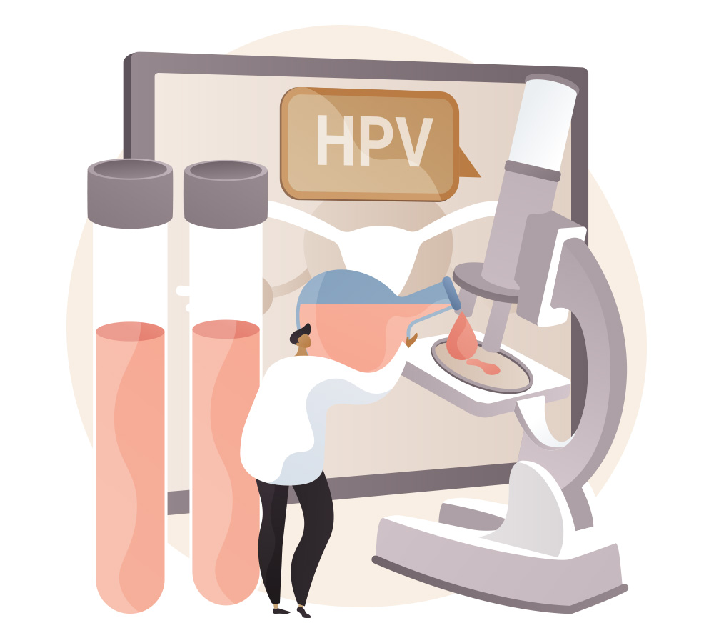
What is the HPV test?
Human papillomaviruses (HPV) are a group of DNA viruses that infect humans. About 200 different viral genotypes of HPV have been described. Of these, 14 have been categorized as high-risk viruses (16,18, 31, 33, 35, 39, 45, 51, 52, 56, 58, 59, 66 and 68) because of their widely known association with the development of premalignant lesions and cervical cancer.
Because of this, the detection of high-risk HPV through the application of molecular biology techniques, specifically real-time polymerase chain reaction (RT-PCR) in cervical cytology samples, is used as an approved and recommended screening method as part of cervical cancer prevention programs.