Laboratory of Anatomic Pathology of El Hematológico

Por: Dra. Laura Tapia Vela
Médica Patóloga, Jefe de Patología Anatómica, Laboratorio Clínico Hematológico S.A. Medellín, Colombia.
Publicado el 11/05/2022

Anatomic pathology is a specialized branch of medicine that studies the cause, origin and nature of disease through the examination of cells, tissues, organs, body fluids and autopsies. For this, it requires a complex approach that includes clinical history, semiology (signs and symptoms), macroscopic anatomical study, light microscopy, the study of cell ultrastructure and molecular analysis.

Despite having a very broad scope and being of great importance in comprehensive medical care, it is striking how most people, even health professionals, are unaware or know little of what goes on or is done in an anatomic pathology laboratory (1) and the diagnostic tools it provides for any patient in and out of the hospital setting (2).

That is why, with this text, we want to help to change this perception, making a brief review of the history of the development of anatomic pathology as a discipline and describing in a general way the services it provides and that allow issuing histopathological diagnoses that direct the treating physician towards the most indicated therapeutic attitude.

History of Pathology

The concept of disease and, therefore, of pathology has changed over the years. In ancient times it was linked to magical explanations; gods, spirits and demons being the main responsible for its appearance. Later, with the arrival of Greek culture and, with it, of the philosophy of human thought, the concept of disease changed towards an organic dimension (3).

Hippocrates and, later, Galen, established the humoral theory of disease, which included four humors (or liquids) identified as black bile, yellow bile, phlegm and blood, which should be in perfect balance to avoid any type of affection or disability. This theory, although not correct, sought to explain the disease from a physiological approach (3).

Teoria Humoral

With the Renaissance, the antecedents of pathology as a discipline emerged. Andrew Vesalius, in the 11th century, studied and developed the anatomical concept of the human body, the morphological basis for the emergence of pathology. Ambrose Paré, the father of surgery, in the 16th century, practiced necropsies to know the cause of death of people. Antoni van Leeuwenhoek, around 1668, built the simple light microscope, with which other important figures of the time, such as Marcelo Malphigi, carried out the microscopic study of organs (4).

The development of anatomical pathology as a medical science began in the 19th century with Xavier Bichat, who buried the erroneous theory of humors, which had existed for more than a thousand years. His experience in the practice of necropsies and his explorations of the human body allowed him to demonstrate that it was formed by tissues and that the inflammation of some of these tissues produced symptoms; and, in turn, these manifestations originated depending on the anatomical location where the affection was found (4).

Thus, with Bichat, a rational basis was given to the explanation of the disease, with its anatomical location in the organs and the symptoms suffered, which represented one of the greatest achievements of medicine, and was the pillar for French authors, headed by René Laënnec, to initiate the development of semiology (4).

anatómica en los órganos

In later years, the routine use of the microscope for the observation of diseased organs moved the study of disease to the cellular stage, where the contributions of Rudolf Virchow, Robert Koch and Carl von Rokitansky, prepared the scientific environment for the discoveries of Louis Pasteur in the field of infectious diseases (3).

This, together with the arrival of Joseph Lister’s asepsis and the emergence of anesthesia, gave rise to the development and progress of surgery and, with it, to surgical pathology, which allowed the study through the microscope of the organs or fragments of organs of a living person, surgically intervened, to make a histopathological diagnosis (4).

It is then when pathology is positioned as the sovereign branch of medicine and the histological diagnosis of biopsy, surgical specimen and cytology acquire a higher value in modern clinical and surgical practice (3).

microscopio

The 20th century was a revolutionary era for pathology, marked by the emergence of countless pioneers in surgical pathology who, coupled with the incorporation of more modern techniques such as immunohistochemistry, flow cytometry and molecular biology, specialized pathologic diagnosis. The era of molecular pathology began with the integration of molecular testing into pathology practice, especially for the diagnosis of solid tumors and hematologic malignancies, as part of the improvements in molecular sciences led by the completion of the Human Genome Project in the early 2000s (5).

Pathologists now play a pivotal role in modern medical care, combining specialized molecular testing with conventional methods of pathologic evaluation to provide accurate disease diagnoses and direct correct treatment plans (5).

It is also important to note telepathology as an active part of the digital era of the 21st century, which is based on the ability to analyze pathology samples from long distances through remote transmission of digital images by telecommunication systems. This sharing of knowledge makes it possible to offer higher quality diagnoses, faster and with a positive impact on patient care.

How does the Anatomic Pathology Laboratory work?

In an anatomic pathology laboratory, different and varied types of samples are received to be analyzed through various methods, including macroscopy, light microscopy, flow cytometry, histochemistry, immunohistochemistry and molecular biology, in order to issue integrated and precise histopathological diagnoses. These precede and determine the therapeutic attitude to be taken by the treating physician, which can be surgical or pharmacological, depending on the conditions of each patient (6).

The most common ways in which samples for pathological analysis can be obtained are: a) by means of a biopsy taken with a thick needle (Trucut), which allows the extraction of a portion of tissue, or by fine needle aspiration (FNA) to collect cellular fluid, b) using an endoscope to observe inside the body the affected areas and obtain small samples from them, or c) by means of surgery in which a part of an organ, a complete organ or several of them are removed.

The success of the pathological diagnosis depends on the correct handling and processing of biopsies and surgical specimens from the moment they are taken. For this reason, immediately after obtaining the tissues, they should be immersed in a fixation medium (10% buffered formalin with neutral pH), in an amount of 15 to 20 times their volume, in containers duly marked with the patient’s identification data. The fixation process avoids the action of enzymes and bacteria that decompose the tissues when they are extracted from an organism and, therefore, achieves their preservation in conditions very similar to normal (6).

Transport time is another critical component in sample preservation. Ideally, they should be delivered to the Pathology laboratory the same day of the procedure; this not only improves the timeliness of the process but also allows for adequate fixation times (6). Once in the pathology laboratory, the pathologist performs the evaluation and macroscopic description (weight, size, shape, consistency, color, margins, among others) of the tissues received.

Tissue samples from biopsies and small specimens are then fully processed and analyzed to ensure their proper evaluation. Meanwhile, large specimens usually have representative parts removed and processed, considered sufficient for analysis, as established by international protocols for each type of sample.

The unprocessed residual material is stored in the original container in which the tissue was sent, thus maintaining its correct identification, with a sufficient amount of 10% buffered formalin to ensure adequate fixation and avoid tissue decomposition. This tissue should be kept until the final report is delivered and then discarded by incineration, after separation of the formalin contained in the sample. The formalin is discarded in a special plastic container for hazardous chemical waste (7).

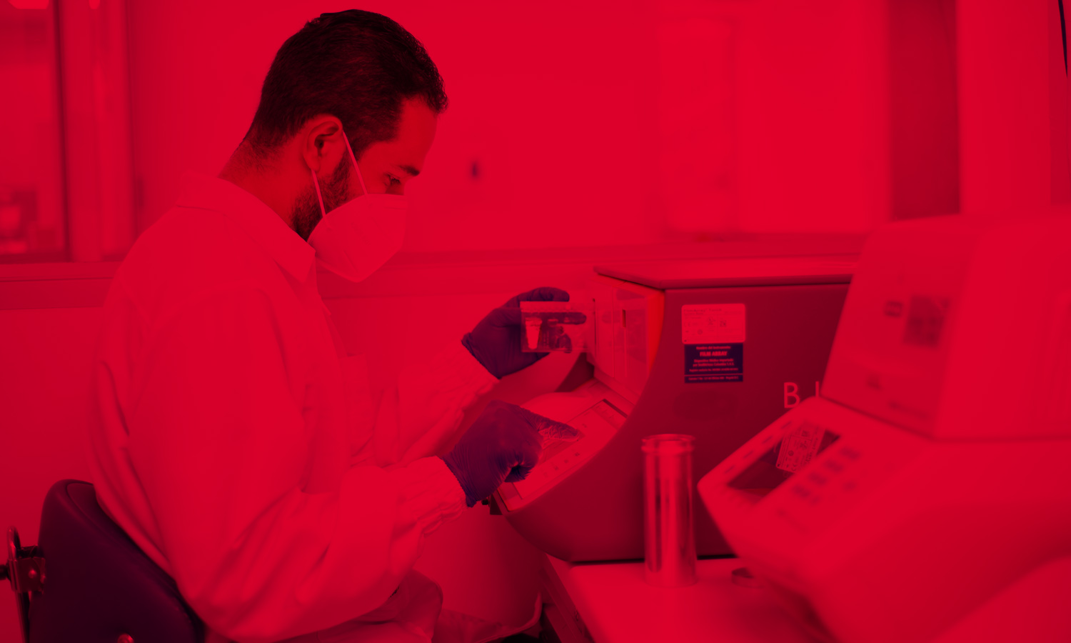
The tissues under analysis are then placed in a tissue processing equipment, where through vacuum systems they pass through formalin, alcohols at different concentrations (70% to 100%), xylol and kerosene, in order to completely fix the sample, dehydrate the tissue and perform the infiltration process with kerosene. All this for its later inclusion in the kerosene blocks in which the sample remains for study.

From each kerosene block, very thin serial cuts (2 to 5 microns) are made, using an instrument called a microtome. These slices are then placed on a slide that is marked with the patient and sample data. Subsequently, the excess kerosene is removed from the slides through the application of heat in an oven or stove, and the routine staining process is performed with hematoxylin-eosin (H/E), which stains the cellular structures and tissues according to their chemical affinity (basophilic or acidophilic) (7).

Once the histological slides with the tissues stained with H/E are available, the pathologist performs the review and analysis through the microscope and interprets the findings in the light of the patient’s clinical data provided by the treating or surgical physician. This allows him to define precise diagnoses (of infections, tumors and other diseases) and to make some recommendations in an integrated pathology report, which will allow him to offer the patient the indicated treatment or management according to his disease.

laminas

It is important to emphasize that the kerosene blocks, histological slides and pathological reports should be archived for approximately 15 years, during which time they should be available for when complementary tests are required, or when research studies of the cases are to be carried out. This is because sometimes, in order to perform a more precise diagnostic approach, or to complement the analysis of the case, it is necessary to add to the pathological study other tests such as special stains, immunohistochemical or molecular tests (ranging from a polymerase chain reaction or PCR, fluorescent in situ hybridization or FISH, to genomic sequencing). All of these are performed from the same kerosene block with the extracted tissue for evaluation that was being processed.

Each of these tests uses different processing and interpretation techniques; for example, immunohistochemistry stains are based on the capacity of tissues to maintain the expression of antigens, which when reacting in an antigen-antibody interaction, by means of an enzymatic reaction marked with color, allow the presence or absence of different markers to be seen and interpreted through the microscope. On the other hand, histochemical stains or special stains use stains that react, according to their physicochemical properties, with specific parts of the tissue, making them stand out, which makes it possible to mark intracellular structures and microorganisms causing infections.

In conclusion, the techniques and processes of pathological analysis allow offering patients and physicians the best and most complete interpretation of cases, focused on the delivery of correct, complete, precise and more clinically useful diagnoses in the diagnostic and therapeutic action, thus contributing to the care and maintenance of the health of the community.

In the Anatomic Pathology Laboratory of Hematológico, we contribute to this medical act with a specialized technical and academic approach, always maintaining the human approach needed to offer a comprehensive and quality diagnostic medicine.


Microbiological diagnosis of chronic bacterial prostatitis

La prostatitis es una de las patologías urológicas más frecuentes y un motivo de consulta común tanto en los hombres menores como en los mayores ...
Leer más →

Liquid-based cervical cytology and Human Papillomavirus

En el Laboratorio Clínico Hematológico nos preocupamos por brindarte las mejores estrategias de tamización para prevenir el cáncer cervicouterino. Actualmente, ofrecemos, en todas nuestras sedes, ...
Leer más →

Linfoma, una mirada desde el laboratorio

El linfoma es un tipo de cáncer del sistema linfático y la sangre que afecta a un grupo específico de los glóbulos blancos, los linfocitos, ...
Leer más →

BiofireⓇ FilmArrayⓇ panels for molecular diagnosis of acute gastrointestinal infections

El panel gastrointestinal (GI) BiofireⓇ FilmArrayⓇes una herramienta molecular que apoya el diagnóstico de infecciones gastrointestinales causadas específicamente por microorganismos patógenos.
Leer más →

Panels for molecular diagnosis of acute respiratory infections

Las infecciones respiratorias agudas (IRA) son, de acuerdo con la Organización Mundial de la Salud (OMS), la principal causa de morbilidad y mortalidad por infección ...
Leer más →

Bibliography

  1. Wilson, M. L. (2018). The Future of Pathology and Laboratory Medicine—Again. American Journal of Clinical Pathology, 150 (2): 93-95. https://doi.org/10.1093/ajcp/aqy058.
  2. Satta, G., & Edmonstone, J. (2018). Consolidation of pathology services in England: Have savings been achieved? BMC Health Services Research, 18(1): 862.  https://doi.org/10.1186/s12913-018-3683-8.
  3. Ariel Gutiérrez Hoyos. (2004). La historia de la Patología en quinientas cincuenta palabras y monosílabos. REV ESP PATOL, 37 (3): 353-354.
  4. Cesar Augusto Giraldo. (2011). El nacimiento de la patología y su desarrollo en la región. Rev Ces Med, 25: 203-211.

¡Hola!
Nos gustaría conocer tu opinión sobre la experiencia que viviste con nuestros servicios.

Encuesta

1. ¿Cómo evalúas tu experiencia con los servicios prestados por el Laboratorio Clínico Hematológico?

Donde 1 es la más baja calificación y 5 la más alta.
2. ¿Cómo calificaría su experiencia global respecto a los servicios de salud que recibió en el Laboratorio?
3. ¿Recomendaría a sus familiares y amigos los servicios del Laboratorio?

Déjanos tu nombre completo, celular y correo electrónico (Opcional)